Abstract
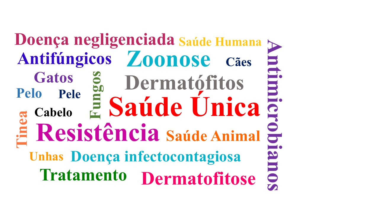
Dermatophytosis is a superficial mycotic infection of keratinized tissues such as hair, nails, and the stratum corneum of the epidermis. In dogs and cats, this disease is commonly caused by dermatophyte fungi of the genera Microsporum, Nannizzia, and Trichophyton, that can affect any patient, with puppies, and elderly, and immunocompromised animals being the most prone. Although it is a highly contagious disease, its mortality is low and in some cases, there can be spontaneous remission. It is estimated that it affects about 4-15% of canines and 20% of felines, being the most common fungal infection in the mentioned species. In addition, dermatophytosis is an anthropozoonosis that affects about 40% of the human population and is widespread in urban centers. It is known that dogs and cats are important carriers of the disease and that both symptomatic and asymptomatic animals are capable of transmitting the agents to each other and humans. Asymptomatic carriers are of great importance for the dissemination of the zoonosis, due to the lack of information and the fact that they do not present lesions, which increases the risks of exposure due to the close contact between tutors and their animals.
References
1. Cafarchia C, Romito D, Capelli G, Guillot J, Otranto D. Isolation of Microsporum canis from the hair coat of pet dogs and cats belonging to owners diagnosed with M. canis tinea corporis. Vet Dermatol. 2006;17(5):327-31. https://doi.org/10.1111/j.1365-3164.2006.00533.x
2. Viani FC. Dermatófitos. In: Jericó MM, Andrade Neto JP, Kogika MM, organizador. Tratado de Medicina Interna de Cães e Gatos. Rio de Janeiro: Roca; 2015.p.2351-65.
3. Oliveira LMB, Pinheiro AQ, Macedo ITF, Silva ING, Moreira OC, Silva BWL et al. Dermatofitose canina causada pelo fungo antropofílico Trichophyton tonsurans – Relato de caso. RBHSA. 2015;9(1):91-8. https://doi.org/10.5935/1981-2965.20150009
4. Silva SF, Teixeira C, Machado S, Marques L. Kérion celsi: uma complicação rara da Tinea capitis. REVNEC. 2017;26(2):126-8. https://doi.org/10.25753/BirthGrowthMJ.v26.i2.9359
5. Hoog GS, Dukik K, Monod M, Packeu A, Stubbe D, Hendrickx M et al. Toward a novel multilocus phylogenetic taxonomy for the dermatophytes. Mycopathologia. 2017;182:5-31. https://doi.org/10.1007/s11046-016-0073-9
6. Balda AC, Otsuka M, Larsson CE. Ensaio clínico da griseofulvina e da terbinafina na terapia das dermatofitoses em cães e gatos. Ciênc Rural. 2007;37(3):750-4. https://doi.org/10.1590/S0103-84782007000300023
7. Moriello KA, Coyner K, Paterson S, Mignon B. Diagnosis and treatment of dermatophytosis in dogs and cats. Clinical Consensus Guidelines of the World Association for Veterinary Dermatology. Vet Dermatol. 2017;28(3):266-8. https://doi.org/10.1111/vde.12440
8. Rossi CN, Zanette MF. Dermatofitose em cães. In: Costa MT, Dagnone AS, organizador. Doenças Infecciosas na Rotina de Cães e Gatos no Brasil. Curitiba: Medvep; 2018.p. 303-34.
9. Terreni AA, Gregg Jr WB, Morris PR, Disalvo AF. Epidermophyton floccosum infection in a dog from the United States. Sabouraudia. 1985;23(2):141-2. https://doi.org/10.1080/00362178585380231
10. Maciel AS, Viana JA. Dermatofitose em cães e gatos – primeira parte. Clín. Vet. 2005;56:48-56.
11. Andrade V, Rossi AM. Dermatofitose em animais de companhia e sua importância para a Saúde Pública – Revisão de Literatura. RBHSA. 2019;13(1):142-55. https://doi.org/10.5935/1981-2965.20190011
12. Peres NTA, Rossi A, Maranhão FCA, Martinez-Rossi NM. Dermatófitos: interação patógeno hospedeiro e resistência a antifúngicos. An Bras Dermatol. 2010;85(5):657-67. https://doi.org/10.1590/S0365-05962010000500009
13. Nweze EI. Dermatophytoses in domesticated animals. Rev Inst Med Trop. 2011;53(2)94-9. https://doi.org/10.1590/S0036-46652011000200007
14. Neves JJA, Paulino AO, Vieira RG, Nishida EK, Coutinho SDA. The presence of dermatophytes in infected pets and their household environment. Arq Bras Med Vet Zootec. 2018;70(6):1747-53. https://doi.org/10.1590/1678-4162-9660
15. Balda AC, Larsson CE, Otsuka M, Gambale W. Estudo retrospectivo de casuística das dermatofitoses em cães e gatos atendidos no Serviço de Dermatologia da Faculdade de Medicina Veterinária e Zootecnia da Universidade de São Paulo. Acta Scientiae Vet. 2004;32(2):133-40. https://doi.org/10.22456/1679-9216.16835
16. Moriello KA, Newbury S. Recommendations for the management and treatment of dermatophytosis in animal shelters. Vet Clin North Am Small Anim. 2006;36:89-114. https://doi.org/10.1016/j.cvsm.2005.09.006
17. Morrow LD. Management of feline dermatophytosis in the rescue shelter environment. Companion Anim. 2016;21(11):634-9. https://doi.org/10.12968/coan.2016.21.11.634
18. De Tar LG, Dubrovsky V, Scarlett JM. Descriptive epidemiology and test characteristics of cats diagnosed with Microsporum canis dermatophytosis in a Northwestern US animal shelter. J Feline Med Surg. 2019;21(12):1198-205. https://doi.org/10.1177/1098612X19825519
19. Gambale W, Larsson CE, Moritami MM, Corrêa B, Paula CR. Dermatophytes and other fungi of the haircoat of cats without dematophytosis in the city of Sao Paulo, Brazil. Feline Pract. 1993;21(3):29-33.
20. Farias MR, Condas LAZ, Ramalho F, Bier D, Muro MD, Pimpão CT. Avaliação do estado de carreador assintomático de fungos dermatofíticos em felinos (Felis catus – linnaeus, 1793) destinados à doação em centros de controle de zoonoses e sociedades protetoras de animais. Vet. zootec. 2011;18(2):306-12. Disponível em: https://rvz.emnuvens.com.br/rvz/article/view/1134
21. DeBoer DJ, Moriello KA. Development of an experimental model of Microsporum canis infection in cats. Vet Microbiol. 1994;42(4):289-95. https://doi.org/10.1016/0378-1135(94)90060-4
22. Newbury S, Moriello KA. Feline dermatophytosis: Steps for investigation of a suspected shelter outbreak. J Feline Med Surg. 2014;16;(5):407-18. https://doi.org/10.1177/1098612X14530213
23. Polak KC, Levy JK, Crawford PC, Leutenegger CM, Moriello KA. Infectious diseases in large-scale cat hoarding investigations. Vet J. 2014;201(2):189-95. https://doi.org/10.1016/j.tvjl.2014.05.020
24. Nitta CY, Daniel AGT, Taborda CP, Santana AE, Larsson CE. Isolation of dermatophytes from the hair coat of healthy persian cats without skin lesions from commercial catteries located in São Paulo metropolitan area, Brazil. Acta Scientiae Vet. 2016;44:1421. https://doi.org/10.22456/1679-9216.81298
25. Scott DW, Paradis M. A survey of canine and feline skin disorders seen in a university practice: Small Animal Clinic, University of Montréal, Saint-Hyacinthe, Québec (1987-1988). Can Vet J. 1990;31(12):830-835. Dsiponível em: https://www.ncbi.nlm.nih.gov/pmc/articles/PMC1480900/
26. Cabañes FJ, Abarca ML, Bragulat MR. Dermatophytes isolated from domestic animals in Barcelona, Spain. Mycopathologia. 1997;137:107-13. https://doi.org/10.1023/A:1006867413987
27. Cafarchia C, Romito D, Sasanelli M, Lia R, Capelli G, Otranto D. The epidemiology of canine and feline dermatophytoses in southern Italy. Mycoses. 2004;47:508-13. https://doi.org/10.1111/j.1439-0507.2004.01055.x
28. Murmu S, Debnath C, Pramanik AK, Mitra T, Jana S, Dey S et al. Detection and characterization of zoonotic dermatophytes from dogs and cats in and around Kolkata. Vet World. 2015;8(9):1078-82. https://doi.org/10.14202/vetworld.2015.1078-1082
29. Moriello K. Dermatophytosis in cats and dogs: a practical guide to diagnosis and treatment. In Pract. 2019;41:138-47. https://doi.org/10.1136/inp.l1539
30. Degreef H. Clinical forms of dermatophytosis (ringworm infection). Mycopathologia. 2008;166(5-6):257-65. https://doi.org/10.1007/s11046-008-9101-8
31. Vermout S, Tabart J, Baldo A, Mathy A, Losson B, Mignon B. Pathogenesis of dermatophytosis. Mycopathologia. 2008;166(5-6):267-75. https://doi.org/10.1007/s11046-008-9104-5
32. Sidrim JJC, Meireles TEF, Oliveira LMP, Diógenes MJN. Aspectos clínico laboratoriais das dermatofitoses. In: Sidrim JJC; Rocha MFG, organizador. Micologia médica à luz de autores contemporâneos. Rio de Janeiro: Guanabara Koogan; 2004.p.120-38.
33. Zaitz C. Dermatofitoses. In: Zaitz C, Campbell I, Marques SA, Ruiz LRB, Framil VMS, organizador. Compêndio de micologia médica. Rio de Janeiro: Guanabara Koogan; 2010.p.157-67.
34. Moriello KA. Treatment of dermatophytosis in dogs and cats: review of published studies. Vet Dermatol. 2004;15(2):99-107. https://doi.org/10.1111/j.1365-3164.2004.00361.x
35. Miller WH, Gfiffin CE, Campbell KL. Miller & Kirk’s Small animal dermatology. St. Louis: Saunders; 2013.
36. Carlotti DN, Bensignor E. Dermatophytosis due to Microsporum persicolor (13 cases) or Microsporum gypseum (20 cases) in dogs. Vet Dermatol. 2002;10(1):17 27. https://doi.org/10.1046/j.1365-3164.1999.00115.x
37. Neves RCSM, Cruz FACS, Lima SR, Torres MM, Dutra V, Sousa VRF. Retrospectiva das dermatofitoses em cães e gatos atendidos no Hospital Veterinário da Universidade Federal de Mato Grosso, nos anos de 2006 a 2008. Ciênc Rural. 2011;41(8):1405-10. https://doi.org/10.1590/S010384782011000800017
38. Moriello KA. Feline dermatophytosis: Aspects pertinent to disease management in single and multiple cat situations. J Feline Med Surg. 2014;16(5):419-31. https://doi.org/10.1177/1098612X14530215
39. Budgin JB. Feline dermatophytosis: an update on diagnosis and treatment. Full circle fórum. 2011;1(7)15-20.
40. Waller SB, Reis-Gomes A, Cabana AL, Faria RO, Meireles MCA, Mello JRB. Microsporose canina e humana – um relato de caso zoonótico. Sci Anim Health. 2014;2(2):137-46. https://doi.org/10.15210/sah.v2i2.4129
41. Ferreira RR, Machado MLS, Spanamberg A, Ferreiro L. Quérion causado por Microsporum gypseum em um cão. Acta Sci Vet. 2006;34(2):179-82. Disponível em: https://lume.ufrgs.br/bitstream/handle/10183/20309/000590148.pdf?sequence=1&isAllowed=y
42. Cornegliani L, Persico P, Colombo S. Canine nodular dermatophytosis (kerion): 23 cases. Vet Dermatol. 2009;20(3):185-90. https://doi.org/10.1111/j.1365-3164.2009.00749.x
43. Reis-Gomes A, Madrid IM, Matos CB, Telles AJ, Waller SB, Nobre MO et al. Dermatopatias fúngicas: aspectos clínicos, diagnósticos e terapêuticos. Acta Vet. Brasilica. 2012;6(4):272-84.
44. Romano C, Valenti L, Barbara R. Dermatophytes isolated from asymptomatic stray cats. Mycoses.1997;40:471-72. https://doi.org/10.1111/j.1439-0507.1997.tb00187.x
45. Boyanowski KJ, Ihrke PJ, Moriello KA, Kass PH. Isolation of fungal flora from the hair coats of shelter cats in the Pacific coastal USA. Vet Dermatol. 2000;11(2):143-50. https://doi.org/10.1046/j.1365-3164.2000.00161.x
46. Bier D, Farias MR, Muro MD, Son LMF, Carvalho VO, Pimpão CT. Isolamento de dermatófitos de pelo de cães e gatos pertencentes a proprietários com diagnóstico de dermatofitose. Arch Vet Sci.2013;18(1):1-8. https://doi.org/10.5380/avs.v18i1.25980
47. Chermette R, Ferreiro L, Guillot J. Dermatophytoses in animals Mycopathologia. 2008;166(5-6):385-405. https://doi.org/10.1007/s11046-008-9102-7
48. Beber MC, Breunig JA. Prurido em região frontal da cabeça. Rev Epidemiol. Control Infect. 2012;2(1):24-5. https://doi.org/10.17058/reci.v2i1.2476
49. Rouzaud C, Hay R, Chosidow O, Dupin N, Puel A, Lortholary O et al. Severe dermatophytosis and acquired or innate immunodeficiency: A review. J Fungi. 2016;2(1):4. https://doi.org/10.3390/jof2010004
50. Kim SH, Jo IH, Kang J, Joo SY, Choi J. Dermatophyte abscesses caused by Trichophyton rubrum in a patient without pre-existing superficial dermatophytosis: A case report. BMC Infec Dis. 2016;16(1):298. https://doi.org/10.1186/s12879-016-1631-y
51. Scott DW, Miller WH, Griffin CE. Muller & Kirk’s. Small Animal Dermatology. California: Saunders; 2001.
52. Roehe C. Gatos portadores de dermatofitos na região sul metropolitana de Porto Alegre – RS, Brasil [dissertação de mestrado]. Porto Alegre (RS): Universidade Federal do Rio Grande do Sul; 2014.
53. Taplin D, Allen AM, Mertz PM. Experience with a new indicator medium for the isolation of dermatophyte fungi. In: Proceedings of the International Symposium on Mycoses. Washington, DC: Pan American Health Organization; 1970.p.55-8.
54. Guillot J, Latié L, Deville M, Halos L, Chermette R. Evaluation of the dermatophyte test medium RapidVet-D. Vet Dermatol. 2001;12(3):123-7. https://doi.org/10.1046/j.1365-3164.2001.00217.x
55. Gondim ALCL, Araújo AKL. Aspectos clínicos, diagnósticos e terapêuticos da dermatofitose em cães e gatos e sua importância como zoonose. Rev Bras de Edu e Saude. 2020;10(1):86-94. https://doi.org/10.18378/rebes.v10i1.7548
56. Gomes JMF. Caracterização dos dermatófitos e leveduras isolados de lesões sugestivas de dermatomicoses em cães [dissertação de mestrado]. Fortaleza (CE): Universidade Estadual do Ceará; 2004.
57. Costa FVA. Determinação da variabilidade genotípica entre isolados de Microsporum canis [tese doutorado]. Porto Alegre (RS): Universidade Federal do Rio Grande do Sul; 2010.
58. Moriello KA. Diagnostic techniques for dermatophytosis. Clin Tech Small Anim Pract. 2001;16(4):219-24. https://doi.org/10.1053/svms.2001.27597
59. Moriello KA, Deboer D. Dermatofitose. In: Greene CE, organizador. Doenças Infecciosas em Cães e Gatos. Rio de Janeiro: Roca; 2015.p.1294-323.
60. Quinn PJ, Markey BK, Carter ME, Donnelly WJC, Leonard FC, Maguire D. Microbiologia veterinária e doenças infecciosas. Porto Alegre: Artemed; 2005.
61. Chengappa MM, Pohlman LM. Dermatófitos. In: Mcvey DS, Kennedy M, Chengappa MM, organizador. Microbiologia Veterinária. Rio de Janeiro: Guanabara Koogan; 2016.p.482-90.
62. Borba LA. Coloração de esporos em pelos na dermatofitose e comparação de técnicas de diagnóstico [dissertação de mestrado]. Curitiba (PR): Universidade Federal do Paraná; 2010.
63. Nardoni S, Franceschi A, Mancianti F. Identification of Microsporum canis from dermatophytic pseudomycetoma in paraffin embedded veterinary specimens using a common PCR protocol. Mycoses. 2007;50(3):215-7. https://doi.org/10.1111/j.1439-0507.2007.01368.x
64. Cafarchia C, Gasser RB, Figueredo LA, Weigl S, Danesi P, Capelli G et al. An improved molecular diagnostic assay for canine and feline dermatophytosis. Med. Mycol. 2013;51(2):136-43. https://doi.org/10.3109/13693786.2012.691995
65. Dabrowska I, Dworecka-Kaszak B, Brillowska-Dabrowska A. The use of a one-step PCR method for the identification of Microsporum canis and Trichophyton mentagrophytes infection of pets. Acta Biochim Pol. 2014;61(2):375-8. Disponível em: https://pubmed.ncbi.nlm.nih.gov/24945136/
66. Peters J, Scott DW, Erb HN, Miller Jr WH. Comparative analysis of canine dermatophytosis and superficial pemphigus for the prevalence of dermatophytes and acantholytic keratinocytes: a histopathological and clinical retrospective study. Vet. Dermatol. 2007;18(4):234-40. https://doi.org/10.1111/j.1365-3164.2007.00599.x
67. Kano R, Edamura K, Yumikura H, Maruyama H, Asano K, Tanaka S et al. Confirmed case of feline mycetoma due to Microsporum canis. Mycoses. 2009;52(1):80-3. https://doi.org/10.1111/j.1439-0507.2008.01518.x
68. Ramadinha RR, Reis RK, Campos SG, Ribeiro SS, Peixoto PV. Lufenuron no tratamento da dermatofitose em cães e gatos. Pesqu Vet Bras. 2010;30(2):132-8. https://doi.org/10.1590/S0100-736X2010000200006
69. Balda AC, Santana AE. Dermatofitose. In: Larsson CE, Lucas R, organizador. Tratado de medicina externa: dermatologia veterinária. São Paulo: Interbook Editora; 2020.p.253-79.
70. Coelho JLG, Saraiva EMS, Mendes RC, Santana WJ. Dermatófito: resistência a antifúngicos. Braz J of Develop. 2020;6(10):74675-86. https://doi.org/10.34117/bjdv6n10-044
71. Fattahi A, Shirvani F, Ayatollahi A, Rezaei-Matehkolaei A, Badali H, Lotfali E et al. Multidrug-resistant Trichophyton mentagrophytes genotype VIII in an Iranian family with generalized dermatophytosis: report of four cases and review of literature. Int J Dermatol. 2021;60(6):686-92. https://doi.org/10.1111/ijd.15226
72. Singh A, Masih A, Monroy-Nieto J, Singh PK, Bowers J, Travis J et al. A unique multidrug-resistant clonal Trichophyton population distinct from Trichophyton mentagrophytes/Trichophyton interdigitale complex causing an ongoing alarming dermatophytosis outbreak in India: Genomic insights and resistance profile. Fungal Genet Biol, 2019;133:103266. https://doi.org/10.1016/j.fgb.2019.103266
73. Hsiao YH, Chen C, Han HS, Kano R. The first report of terbinafine resistance Microsporum canis from a cat. J Vet Med Sci. 2018;80(6):898-900. https://doi.org/10.1292/jvms.17-0680
74. Martinez-Rossi NM, Bitencourt TA, Peres NTA, Lang EAS, Gomes EV, Quaresemin NR et al. Dermatophyte resistance to antifungal drugs: mechanisms and prospectus. Front Microbiol. 2018;9:1108. https://doi.org/10.3389/fmicb.2018.01108
75. Khurana A, Sardana K, Chowdhary A. Antifungal resistance in dermatophytes: Recent trends and therapeutic implications. Fungal Genet Biol. 2019;132:103255. https://doi.org/10.1016/j.fgb.2019.103255
76. Espinel-Ingroff A. Standardized disk diffusion method for yeasts. Clin Microbiol Newsl. 2007;29(13):97-100. https://doi.org/10.1016/j.clinmicnews.2007.06.001
77. Alpun G, Ozgur NY. Mycological examination of microsporum canis infection in suspected dermatophytosis of owned and ownerless cats and its asymptomatic carriage. J Anim Vet Adv. 2009;8(4):803-6.
78. Deboer DJ, Moriello KA. The immune response to Microsporum canis induced by a fungal cell wall vaccine. Vet Dermatol. 1994;5(2):47-55. https://doi.org/10.1111/j.1365-3164.1994.tb00011.x
79. Wawrzkiewicz K, Sadzikowski Z, Ziólkowska G, Wawrzkiewicz J. Inactivated vaccine against Microsporum canis infection in cats. Med Weter. 2000;56(4):245-50.

This work is licensed under a Creative Commons Attribution 4.0 International License.
Copyright (c) 2025 Breno Henrique Alves, Bruna Carioca de Souza, Gabriela Ribeiro Pedrosa Rotundo, Sávio Tadeu Almeida Junior , Carlos Artur Lopes Leite, Ana Paula Peconick, Geraldo Márcio da Costa
